Transforming Cancer Treatment: Latest Medical Imaging Techniques.

Fighting Cancer Blindly: Why Medical Imaging is the Key to Winning the Battle
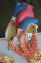
Being diagnosed with cancer can be one of the most frightening experiences in a person’s life. However, what many people don’t realize is that once they receive their diagnosis, they may be fighting the disease blindly without access to medical imaging. Medical imaging, such as CT scans and MRIs, can provide critical information about the location, size, and severity of cancer, helping doctors to make informed decisions about treatment options.
When I was diagnosed with cancer a few years ago, my doctor ordered a series of medical imaging tests to help determine the best course of action. The images revealed the exact location of the tumor, its size, and its proximity to other organs. This information was crucial in deciding which treatment approach to take, as well as the likelihood of successful treatment.
Without medical imaging, doctors may have to rely on guesswork when it comes to treating cancer. This can result in treatments that are either too aggressive, causing unnecessary harm to the patient, or too conservative, allowing the cancer to spread or worsen. Medical imaging can help doctors to make more accurate diagnoses, choose the most effective treatment options, and monitor the progress of treatment over time.
Of course, medical imaging is not without its risks, and it is important for patients to understand the potential benefits and drawbacks before undergoing any tests. Patients should always talk to their doctor about any concerns they may have, and ask questions about the risks, benefits, and alternatives to medical imaging.
In conclusion, medical imaging is a powerful tool in the fight against cancer, and can make all the difference in a patient’s journey to recovery. By providing critical information about the location, size, and severity of cancer, medical imaging can help doctors to make informed decisions about treatment, resulting in better outcomes for patients.
The Reality of Cancer Today: Why We Have Not Won the War
Cancer is a disease that has affected almost everyone in some way or another. Whether it be a loved one, a friend, or a colleague, cancer has the power to impact our lives in profound ways. Despite the advances made in cancer treatment over the years, we have not yet won the war against this disease.
There are several reasons why we have not yet won the war against cancer. One of the main reasons is that cancer is not a single disease, but rather a collection of diseases that are each unique in their own way. Cancer can affect any part of the body, and each type of cancer behaves differently, making it difficult to find a one-size-fits-all treatment.
Another reason why we have not yet won the war against cancer is that cancer cells are extremely adaptable. Cancer cells can evolve and change, making them resistant to treatments that were once effective. This means that even if a treatment works for a while, it may eventually stop working as the cancer cells adapt and become resistant to it.
Additionally, there is still much we do not know about cancer. While we have made significant strides in understanding the disease, there is still much more to be learned. Cancer is a complex disease that involves multiple biological processes, and we are still discovering new information about how it works.
Despite these challenges, there is hope. Advances in medical imaging, such as MRI, PET, and CT scans, have given us powerful tools to detect cancer earlier and more accurately. This means that cancer can be treated before it has a chance to spread, increasing the chances of a successful outcome.
In conclusion, while the fight against cancer is far from over, there is hope. By continuing to invest in research and development, and by leveraging the latest medical imaging technology, we can continue to make progress in the fight against this disease. With continued effort and dedication, we can work towards a future where cancer is no longer a life-threatening disease.
The Story of Ehud: How Brain Cancer Took a Friend’s Life
Cancer is a devastating disease that can strike anyone, anywhere, at any time. Unfortunately, my friend Ehud was not immune to this harsh reality. He was diagnosed with brain cancer, and it changed everything for him.
Ehud was a brilliant and talented man. He was a gifted musician, and he had a passion for life that was infectious. We met in college, and we quickly became close friends. I remember spending countless hours with him, playing music, talking about life, and dreaming about the future.
But one day, everything changed. Ehud started experiencing strange symptoms, like headaches and vision problems. He went to see a doctor, and that’s when he received the devastating news that he had a brain tumor.
Ehud fought the cancer with everything he had. He underwent surgery, chemotherapy, and radiation, but nothing seemed to work. The cancer was too aggressive, and it had already spread to other parts of his body.
Watching Ehud go through this was heartbreaking. He was such a strong and resilient person, but cancer had taken hold of his body, and there was nothing anyone could do to stop it. He passed away a few months after his diagnosis, leaving behind a devastated family, and a group of friends who loved him deeply.
Ehud’s story is a tragic one, but it’s a reminder of the harsh reality of cancer. It’s a disease that can strike anyone, at any time, and it can be incredibly difficult to treat. That’s why it’s so important to continue investing in cancer research, so that we can find better treatments and, ultimately, a cure.
PET scans and the limitations of current cancer detection methods
Detecting cancer in its early stages is crucial for successful treatment and recovery. PET scans are one of the most commonly used imaging techniques for cancer detection. They use a special dye that contains radioactive tracers to highlight areas of abnormal metabolic activity in the body. However, as effective as they may be, PET scans do have their limitations.
One of the main limitations of PET scans is their ability to accurately distinguish between cancerous and non-cancerous cells. This is because PET scans detect metabolic activity, which can also occur in normal cells that are actively dividing or repairing themselves. Therefore, PET scans can produce false positives, leading to unnecessary biopsies or treatments, as well as false negatives, where cancer is missed entirely.
Another limitation is the availability and cost of PET scans. They are not readily available in all healthcare facilities, and their cost can be prohibitive for many patients. In addition, PET scans expose patients to ionizing radiation, which can be harmful over time.
Despite these limitations, PET scans remain a valuable tool in cancer detection and treatment. However, continued research and development of new imaging technologies are necessary to improve their accuracy and accessibility.
The Vital Role of Complete Cancer Cell Removal in Surgery
Cancer surgery is one of the most common treatment options for cancer patients. Although advancements in medical science have made cancer surgery safer and more effective, complete removal of cancer cells during surgery remains a crucial factor that can make or break the success of the surgery.
During surgery, surgeons try to remove as much of the cancerous tissue as possible while preserving the healthy tissues. However, it is often challenging to determine the exact extent of the cancerous tissue, and if any malignant cells are left behind, they can potentially grow and spread to other parts of the body, leading to recurrence.
Therefore, it is important for the surgeon to remove the entire tumor, including its margins, as well as any nearby lymph nodes that may contain cancer cells. This is why surgeons use techniques like frozen section analysis and intraoperative imaging to help guide their decision-making during the surgery.
The frozen section analysis allows the surgeon to evaluate the tissue margins and determine if any malignant cells are still present in the removed tissue. Intraoperative imaging, such as fluorescent-guided surgery, uses fluorescent markers that bind to cancer cells and highlight their location, making it easier for the surgeon to remove them completely.
By ensuring the complete removal of cancer cells during surgery, patients have a better chance of long-term survival and reduced risk of cancer recurrence. Therefore, it is important for patients to discuss their surgery options thoroughly with their medical team and understand the importance of complete cancer cell removal in their treatment plan.
Using Gold Nanoparticles to Transform Cancer Surgery
Gold nanoparticles have shown great potential in improving cancer surgery by making it easier to remove all cancerous cells during the procedure. These tiny particles are only a few nanometers in size, but they have unique properties that make them highly effective in cancer treatment.
When gold nanoparticles are introduced into the body, they can be targeted to specific cancer cells using antibodies or other molecules. Once the particles have attached to the cancer cells, they can be heated using a laser to destroy the cells. This process is known as photothermal therapy, and it can be used to selectively kill cancer cells while leaving healthy cells untouched.
In addition to their use in photothermal therapy, gold nanoparticles can also be used to enhance the imaging of cancerous tissues. When gold nanoparticles are injected into the body, they can be detected using imaging techniques such as CT scans, allowing doctors to more accurately locate cancerous tissues during surgery.
Overall, the use of gold nanoparticles in cancer treatment holds great promise for improving the accuracy and effectiveness of cancer surgery. While more research is needed to fully understand their potential, these tiny particles have already shown great promise in the fight against cancer.
A new era in medical imaging: The ability to see every cell in the body
Medical imaging is an essential tool in the fight against cancer. The ability to visualize the tumor, its size, and location in the body is crucial in deciding the best treatment course. However, current imaging techniques have limitations, and sometimes cancer cells can be missed, leading to incomplete removal during surgery.
Advances in medical imaging are transforming cancer treatment. New technologies, such as single-cell imaging and gold nanoparticles, are pushing the boundaries of what is possible. Scientists are now able to see every cell in the body and detect even the smallest tumors.
This breakthrough in imaging is critical in the fight against cancer. The more precise we can be in detecting and removing cancer cells, the better the chances of a successful outcome. With this new technology, we can ensure that every cancer cell is removed during surgery, greatly reducing the risk of recurrence.
In conclusion, medical imaging has come a long way in recent years, and this new era in imaging technology brings us one step closer to winning the war on cancer. By visualizing every cell in the body, we can detect and remove cancer cells more accurately than ever before, giving patients a better chance of beating the disease.
Asking Cancer Cells Real Questions: The Potential for Groundbreaking Insights
One of the most significant challenges in cancer research is getting real insights into how cancer cells behave. Traditional methods rely on studying cells outside of the body or in animal models, which do not always accurately reflect how cancer cells behave in humans.
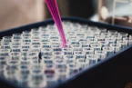
Recent advances in medical imaging have allowed researchers to ask cancer cells real questions, providing a unique opportunity for groundbreaking insights. This new technology uses tiny, flexible needles to extract cancer cells directly from the body and analyze them in a lab setting.
By studying cancer cells in this way, researchers can gain insights into how the cells interact with one another, how they respond to different treatments, and what makes them grow and spread. This knowledge can then be used to develop more effective treatments and personalized therapies that target specific types of cancer cells.
The potential impact of this technology is enormous. By being able to ask cancer cells real questions, we can unlock new insights that were previously unimaginable. It opens up a whole new world of possibilities in the fight against cancer, and we are excited to see what the future holds.
Conclusion
Cancer is a complex and devastating disease that affects millions of people worldwide. While significant progress has been made in cancer research and treatment, there is still much work to be done to improve outcomes for patients. Medical imaging, such as PET scans and gold nanoparticles, have shown great potential in improving cancer detection and surgery. The ability to see every cell in the body and ask cancer cells real questions through new imaging technologies provides unprecedented opportunities for groundbreaking insights.
However, it is important to recognize that there are limitations and challenges that must be overcome in the fight against cancer. Every patient’s journey is unique, and it is crucial to approach cancer treatment and care with compassion, empathy, and a commitment to personalized medicine. Together, researchers, clinicians, patients, and their loved ones can work towards a future where cancer is no longer a devastating diagnosis, but a treatable and even curable condition.